PGD is carried out as part of in vitro fertilization (IVF or “test tube baby”) treatment only. After follicle stimulating hormone treatment several oocytes, or eggs, are retrieved from the woman. The oocytes are then fertilized in vitro, outside the body. The embryos developing from the zygotes are generally implanted in the uterus on day 3 or day 5 of development. In the course of the procedure it is possible to take samples for genetic testing. In the case of oocytes and zygotes (i.e. fertilized eggs) the test is aimed at identifying a genetic change in the so-called polar body.
- Polar body biopsy: In the case of oocytes and zygotes (i.e. fertilized eggs) the test is aimed at identifying a genetic change in the so-called polar body. The drawback of the procedure is that it provides information only about the mother’s genome, and also that only a single cell can be tested.
- Blastomere biopsy: A blastomere (i.e. a sample of two or three cells) of the three-day-old embryo is tested. Implantation will take place on day 5. The drawback of this method is that only one or two cells are analysed and thus the genetic opinion does not cover of all of the embryos, and also there is a relatively high rate (5-10 %) of false negative or false positive results.
- Trophectoderm biopsy: On day 5, at the blastocyst stage, a small portion of the so-called trophectoderm layer consisting of 20-50 cells is removed for testing. This is the most reliable procedure, so in future it will probably become the most widely used method. Its drawback is that the embryos must be frozen and implantation can take place only in the next cycle.
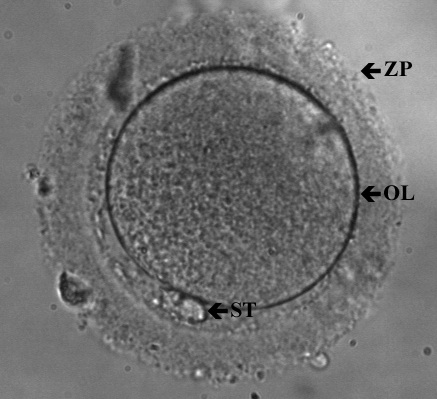
Oocyte in metaphase II of the meiotic process
Gametes are produced by meiosis. Meiosis takes place in two stages. Meiosis of the oocyte ready to be fertilized arrests at meiosis II, the second phase, and in a normal case it is only completed after fertilization as a result of activation triggered by the sperm.
ZP: zona pellucida (the outer layer of the oocyte)
OL: oolemma (cytoplasm membrane of the oocyte)
ST: polar body (the cell resulting from the first meiotic phase)
In the course of the procedure the oocyte or the embryo is placed in a special medium. A tiny incision or hole is made on the zona pellucida, generally by means of laser, and the cell or cells to be tested are retrieved through the hole with a micropipette, a very thin glass capillary.
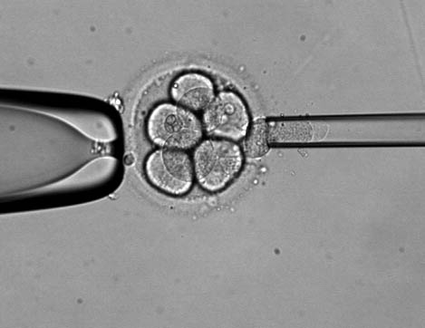
Blastomere biopsy
On day 3 the pre-embryos are generally at the 5-10 cells stage. At this stage one or two cells (blastomere) are removed by means of a micromanipulator from the embryo for the purpose of pre-implantation genetic diagnostics.
After preparation the sample is taken to the genetic lab, where various molecular biochemical test methods are applied (FISH, PCR, CGH) depending on the purpose of the test and the disease to be detected.